Andhra Pradesh BIEAP AP Inter 1st Year Botany Study Material 12th Lesson Histology and Anatomy of Flowering Plants Textbook Questions and Answers.
AP Inter 1st Year Botany Study Material 12th Lesson Histology and Anatomy of Flowering Plants
Very Short Answer Questions
Question 1.
The transverse section of a plant material shows the following anatomical features.
a) The vascular bundles are conjoint, scattered, and surrounded by sclerenchyma matous bundle sheaths.
b) Phloem parenchyma is absent. What will you identify it as?
Answer:
Monocot Stem.
Question 2.
Why are xylem and phloem called complex tissues?
Answer:
Xylem and phloem are permanent tissues having more than one type of cells and work together. So-called complex tissues.
Question 3.
How is the study of plant anatomy useful to us?
Answer:
First of all the study of plant Anatomy helps us understand the way a plant functions, carrying out its routine activities like transpiration, photosynthesis, growth and repair. Second, it helps botanists and agriculture scientists to understand the disease and cure for the plants. Anatomy is way to understand the larger system of Ecology on this planet.
Question 4.
Protoxylem is the first formed xylem. If the protoxylem lies rodialy next to pholem what kind of arrangement of xylem would you call it? Where do you find it?
Answer:
Radial vascular Bundle. They are found in Roots.
Question 5.
What is the function of phloem parenchyma?
Answer:
Phloem parenchyma stores food material and other substances like resins, latex and mucilage.
![]()
Question 6.
a) What is present on the surface of the leaves which helps the plant to prevent loss of water but is absent in roots?
b) What is the epidermal cell modification in plants which prevents water loss?
Answer:
(a) Cuticle
(b) Trichomes.
Question 7.
Which part of the plant would show the following?
(a) Radical vascular bundle (b) Polyarch xylem (c)Well developed pith (d) Exarch xylem
Answer:
a) Root
b) Monocot Root
c) Monocot Root
d) Roots
Question 8.
What, are the cells that make the leaves curl in plants during water stress? Give an example.
Answer:
Bulliform cells. Ex : Monocot leaf (grass).
Question 9.
What constitutes the Vascular combial ring?
Answer:
Intrafascicular cambium + Interfascicular cambium constitutes cambial ring.
Question 10.
Give one basic functional difference between phellogen and phelloderm.
Answer:
Phellogen is also called cork cambium which appears in cortex and produces cork and phelloderm. The cells in the phellogen are thin walled, rectangular.
Phelloderm :
The cells which are formed towards inside from phellogen constitutes phelloderm or secondary cortex. The cells are parenchymatous.
Question 11.
If one debarks a tree, what parts of the plant are removed?
Answer:
Periderm and secondary phloem.
Short Answer Type Questions
Question 1.
State the location and function of different types of meristems.
Answer:
Based on the position, meristems are classified into three types.
A) Apical Meristems :
The meristems that are present at the tip of the root and at the tip of the stem or branches are called Apical Meristems. They help in the linear growth of the plant body.
B) Intercalary Meristems :
The Meristems that are present in between mature tissues are known as Intercalary Meristems. They occur in grasses. They also contribute to the formation of the primary plant body.
C) Lateral Meristems :
The meristems that occur in the mature regions of roots and shoots of many plants are called lateral meristems. They help in increase in thickness of the plant organs (Root, Stem). Ex : Vascular cambium, cork cambium.
Question 2.
Cut a transverse section of young stem of a plant from your garden and observe it under the microscope. How would you ascertain whether it is a monocot stem or a dicot stem? Give Reasons.
Answer:
The transverse section of a young stem which was observed under Microscope shows some characters to indicate that it is either Monocot or Dicot. They are ;
| Dicot stem | Monocot Stem |
| 1. Epidermis have trichomes. | 1. Trichomes are absent. |
| 2. Hypodermis is collenchymatous. | 2. Hypodermis is sclerenchymatous. |
| 3. General cortex and endodermis are present. | 3. General cortex and endodermis are absent. |
| 4. Ground tissue is absent. | 4. Ground tissue present. |
| 5. Vascular bundles are limited in number and are arranged in a ring (Eustele). | 5. Vascular bundles are numerous, scattered, irregularly in the ground tissue (attactostele). |
| 6. Vascular Bundles are top shaped. | 6. Vascular bundles are oval shaped. |
| 7. Vascular Bundles are collateral, conjoint, open type. | 7. Vascular bundles are collateral, conjoint and closed type. |
| 8. Xylem vessels are more. | 8. Xylem vessels are few. |
| 9. Protoxylem lacuna is absent. | 9. Protoxylem lacuna is present. |
| 10. Xylem vessels are in a row. | 10. Xylem vessels are in the form of ‘V’ shape. |
| 11. Medulla, Medullary rays are present. | 11. Medulla, Medullary rays are absent. |
| 12. Phloem parenchyma is present. | 12. Phloem parenchyma is absent. |
With the above differences. We can observe the stem, whether it belongs to Dicot or Monocot stem.
![]()
Question 3.
What is periderm? How does periderm formation take place in the dicot stems?
Answer:
Phellogen, phellem and phelloderm are collectively known as “Periderm”.
Periderm :
Due to the formation secondary vascular tissues inside the stele, a pressure is cortex on the epidermis causing it to rupture. In the mean while, a secondary protective layer is formed called ‘periderm’ from the cortex. The secondary growth in the cortex begins with the appearence of a meristematic layer of cells from the middle part of the cortex.
This is called “Phellogen or cork cambium”. The cells of the phellogen divide periclinally and cuts of new cells on either side. The cells produced towards outside are called cork cells or phellem and the cells produced towards inside are called “secondary cortex cells or phelloderm”. The phellogen (cork cambium), phellum (cork) and phelloderm (secondary cortex) together constitute “periderm”.
Question 4.
A transverse section of the trunk of a tree, shows concentric rings which are known as annual rings. How are these rings formed? What is the significance of these rings?
Answer:
Annual rings :
In temperate regions and cold regions the activity of the cambium is influenced by the seasonal variations. During the favourable season, i.e., in spring, when more leaves and flowers are formed, the plant requires large amounts of water and mineral salts. Hence the wood formed in this period shows more number of xylem vessels with wider lumens.
This is known as spring wood or early wood. The colour of this wood is light. During the unfavourable season i.e., in autumn, the plants are less active and do not require more water and mineral salts. Hence the wood produced in this period shows less number of xylem vessely with narrow lumens. This is known as autumn wood or late wood. It is dark coloured. In this way two types of secondary xylem (wood) are produced in one year. They appear in the form of dark and light coloured circles alternately in a mature tree trunk. These are called annual rings or growth rings or seasonal rings.
By counting the number of annual rings, the approximate age of trees can be estimated. This branch of science is known as “dendrochronology or growth ring analysis”. For Ex : The age of sequoid dendron, presently growing in America, is estimated to be about 3500 years. In tropical countries like India, annual rings do not appear clearly, as the seasonal variations are not sharp. Hence these are called growth marks.
Question 5.
What is the difference between lenticels and stomata?
Answer:
| Lenticels | Stomata |
| 1. Lenticels are portions of the periderm (Bark) with numerous Intercellular spaces. | 1. Stomata occur in the Epidermis of leaves and younger stems. |
| 2. Lenticels does not have Guard cells. | 2. Stoma has Guard cells. |
| 3. Lenticels are present on the outer layer of woody or Hard stem. | 3. Stoma are present on the lower surface of the leaf. |
| 4. They are used for removal of waste. | 4. They are involved in gaseous exchange, removal of extra water and waste. |
Question 6.
Write the precise function of
(a) Sieve tube (b) Interfasicular cambium (c) Collenchyma (d) Sclerenchyma.
Answer:
a) Sieve tube :
They are the more advanced type of conducting cells are and found in phloem of Angiosperms. They are elongated cells, arranged end to end and functioning to conduct food materials through out the plant. They are a nucleate living cells.
b) Interfasicular cambium :
The parenchymatous cells present between vascular Bundles, become meristamatic and form a cambium called Interfascicular cambium.
c) Collenchyma :
- It is a living mechanical tissue cellwall is composed of cellulose, hemicellulose and pectin.
- Collenchyma cells with chloroplasts perform photosynthesis.
- They provide mechanical support to the growing parts of the plants such as young stem and petiole of a leaf.
d) Sclerenchyma :
It is Dead mechanical tissue. Cell wall is made up of lignin. Intercellular spaces are absent.
- Fibres are useful in textile and jute industries. ,
- Fibres give mechanical support to the plant parts.
- Sclerids give mechanical support to the plant parts.
Question 7.
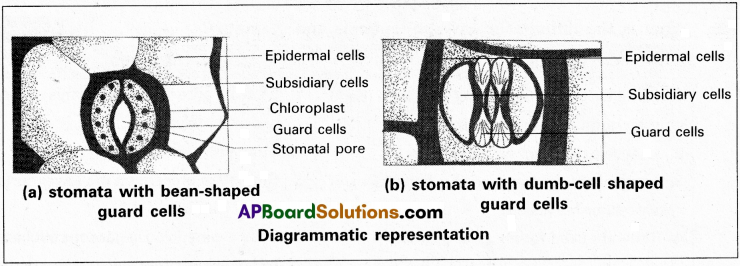
The stomatal pore is guarded by two kidney shaped guard cells. Name the epidermal cells surrounding the guard cells. How does a guard cell differ from an epidermal cell? Use a diagram to illustrate your answer.
Answer:
Stomata are structures present in the epidermis of leaves. Each stomata is composed of two bean shaped cells known as guard cells. The outer walls of guard cells are thin and the inner walls are thick. The guard cells possess chloroplasts and regulate the opening and closing of stomata. Sometimes a few epidermal cells, become specialized in their shape and size are called subsidiary cells. Differences between Guard cell and Epidermal cells.
| Guard cells | Epidermal cells |
| 1. They are bean or kidney shaped. | 1. They are Barrel shaped. |
| 2. They possess chloroplasts. | 2. They lack chloroplasts. |
| 3. They are smaller. | 3. They are bigger. |
| 4. Cell walls of Guard cells are not uniform and Thicker. | 4. Epidermal cells are uniformly thin. |
Question 8.
Point out the differences in the anatomy of leaf of peepal (Ficus religiosa) and Maize (Zea mays). Draw the diagrams and label the differences.
Answer:
| Dicot leaf | Monocot leaf |
| 1. Stomata are more in the lower epidermis than in the upper epidermis. | 1. Stomata are equally distributed on both the sides. |
| 2. Bulliform cells are absent. | 2. Bulliform cells are present in the upper epidermis. |
| 3. Mesophyll is differentiated into palisade and spongy tissue. | 3. Mesophyll is undifferentiated. |
| 4. Bundle sheath extensions are generally parenchymatous. | 4. Bundle sheath extensions are generally sclerenchymatous. |
5. They are dark green on the upper surface, less green on the lower surface. (Dorsiventral)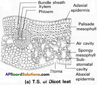 | 5. They are in same colour on both the surfaces. (Isobilateral)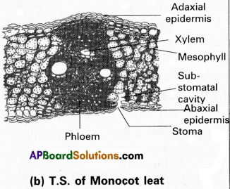 |
Question 9.
Cork cambium forms tissues that form the cork. Do you agree with this statement? Explain.
Answer:
Yes, cork cambium or phellogen is formed during secondary growth of Dicot stem. Cork cambium or phellogen cuts off on both sides. The cells produced towards outerside differentiates into cork or phellem and the inner cells differentiates into secondary cortex or phelloderm. The cork cells are impervious to water due to deposition of suberin in the cell wall.
![]()
Question 10.
Name the three basic tissue systems in the flowering plants. Give the tissue names under each system.
Answer:
In flowering plants, there are three tissue systems are present namely.
- Epidermal tissue system
- Ground tissue system
- Vascular tissue system.
1) Epidermal tissue system :
It consists of epidermis, cuticle, stomata, unicellular hairs and Multicellular trichomes.
2) Ground tissue system :
It consists of simple tissues like parenchyma, collenchyma and sclerenchyma. These cells are present in cortex, pericycle, pith, Medullary rays, Hypodermis, Endodermis layers. In leaves, the ground tissue consist of thin walled chloroplast containing cells called Mesophyll.
3) Vascular tissue system :
It consists of complex tissues, the xylem and the phloem.
Long Answer Type Questions
Question 1.
Explain the process of secondary growth in the stems of woody Angiosperms with the help of schematic diagrams. What is its significance?
Answer:
Formation of cambium ring :
In the primary structure of dicto stem, the stele shows vascular bundles in the form of a ring. Each vascular bundle consists of cambium in between the xylem and phloem. This is called Inter fascicular cambium. In between the vascular bundles, there are medullary rays. From the cells of medullary rays intrafascicular cambium is formed. The Inter fascicular and intrafascicular cambia fuse to form a continuous cambial ring called “vascular cambium”.
Activity of the vascular cambial ring :
The cells of vascular cambium divide repeatedly by periclinal method and produce new cells on both the sides. The cells which are produced outside develop into secondary phloem and those produced to the inner side develop into secondary xylem (wood). Generally more secondary xylem is produced than the secondary phloem. The secondary xylem consists of xylem vessels, tracheids, xylem fibres and xylem parenchyma. The secondary phloem consists of sieve tubes, companion cells, phloem fibres and phloem parenchyma.
In the cambium two types of initiating cells are found. They are 1. Fusiform initials and 2. Ray initials. The fusiform initials give rise to the secondary xylem and the secondary phloem. The ray initials produce phloem rays (bast rays) to the outside and xylem rays (wood rays) to the inside. They are helpful in lateral conduction and storage. They are called secondary medullary rays.
Annual rings :
In temperate regions and cold regions the activity of the Cambium is influenced by the seasonal variations. During the favourable season, i.e., in spring, when more leaves and flowers are formed, the plant requires large amounts of water and mineral salts. Hence the wood formed in this period shows more number of xylem vessels with wider lumens. This is known as spring wood or early wood. The colour .of this wood is light during the unfavourable season ie., in autumn, the plants are less active and do not requirfe more water and mineral salts.
Hence the wood produced in the period shows less number of xylem vessels with narrow lumens. This is knwon as Autumn wood or late wood. It is dark coloured. In this way two types of secondary xylem (wood) are produced in one year. They appear in the form of dark and light coloured circles alternately in a mature tree trunk. These are called Annual rings or growth rings or seasonal rings.
By counting the number of annual rings, the approximate age of trees can be estimated. This branch of science is known as “dendrochronology or growth ring analysisFor example the age of sequoid dendron, presently growing in America, is estimated to be about 3500 years, in tropical countries like India, annual rings do not appear clearly, as the seasonal variations are not sharp. Hence these are called growth marks.
Heart wood and sapwood :
With the increase in the age of the tree, the wood undergoes a number of hysical and chemical changes. The older wood gradually loses water and stores food substances and becomes infilterated with various organic compounds such as oils, gums, resins, tannins, colouring agents and aromatic substances. Hence the older xylem present in the centre appears dark in colour. This is called heart wood or duramen.
It is very hard highly durable. Heart wood cannot conduct water and salts because of the growth of tyloses in the lumens of xylem vessels. The heart wood gives mechanical strength to the tree.
The newly formed secondary xylem is found in the peripheral part of the tree trunk. This is called sapwood or alburnum. It is light in colour and is active in conducting water, mineral salts and storage of food materials. As time passes on, the sap wood gradually changes into heart wood. Hence the sap wood remains uniformly thick.
Periderm :
As the secondary xylem and secondary phloem are formed inside the stele, a pressure is exerted on the epidermis, causing its rupture. Mean while a secondary protective layer formed from the middle or inner part of the cortex become meristematic and acts as phellogen or cork cambium. These cells divide periclinally and cuts of new cells towards outside called cork or phellem and towards inside called secondary cortex or phelloderm. The phellogen, phellem and phelloderm together constitute periderm.
At certain regions, the phellogen cuts off closely arranged parenchymatous cells on the outer side instead of cork cells, called complementary cells. These cells soon rupture the epidermis forming a lens-shaped oepnings called lenticels. They permit the exchange of gases between the outer atmosphere and the internal tissues.
Question 2.
Draw illustrations to bring out the anatomical differences between
a) Monocot root and Dicot root
b) Monocot stem and Dicot stem.
Answer:
a) Monocot root and Dicot root
| Monocot Root | Dicot Root |
| 1. Cortex is relatively bigger. | 1. Cortex is smaller. |
| 2. Pericycle is often multilayered. | 2. Pericycle is single layered. |
| 3. Pericycle produces only lateral roots. | 3. Pericycle gives rise to lateral roots and also produces vascular cambium during secondary growth. |
| 4. Vascular bundles are more than six in number. | 4. Vascular bundles range from two to six in number. |
| 5. Xylem is ployarch. | 5. Xylem is monarch to tetrarch. |
| 6. Medulla is very big. | 6. Medulla is very small or absent. |
| 7. Secondary growth is absent. | 7. Secondary growth occurs. |
b) Monocot stem and Dicot stem.
| Monocot Stem | Dicot Stem |
| 1. Trichomes are absent. | 1. Trichomes are present. |
| 2. Hypodermis is made up of sclerenchymatous cells. | 2. Hypodermis is made up of collenchymatous cells. |
| 3. Endodermis and pericycle are absent. | 3. Endodermis and pericycle are present. |
| 4. General cortex is absent. | 4. General cortex is present. |
| 5. Ground tissue is present. | 5. Ground tissue is absent. |
| 6. Vascular bundles are numerous and arranged in a scattered manner (atactostele). | 6. Vascular bundles are few in number and arranged as a circular ring (eustele). |
| 7. Vascular bundle is oval in shape. | 7. Vascular bundle is top shaped or wedge shaped. |
| 8. Vascular bundle is enclosed by fibrous sheath. | 8. Vascular bundle is not enclosed by firbrous sheath. |
| 9. Vascular bundle is closed. | 9. Vascular bundle is open. |
| 10. Xylem vessels are few in number. | 10. Xylem vessels are more in number. |
| 11. Protoxylem lacunae are present. | 11. Protoxylem lacunae are absent. |
| 12. Medulla and medullary rays are absent. | 12. Medulla and medullary rays are present. |
| 13. Pith cavities are present. | 13. Pith cavities are absent. |
| 14. Vessels are in ‘Y’ shape. | 14. Vessels are in serial order. |
| 15. Phloem parenchyma is absent. | 15. Phloem parenchyma is present. |
![]()
Question 3.
What are simple tissues? Describe various types of simple tissues.
Answer:
Tissues which are made up of only one type of cells are called simple tissues. They are of three types. They are parenchyma, Collenchyma and Sclerenchyma.
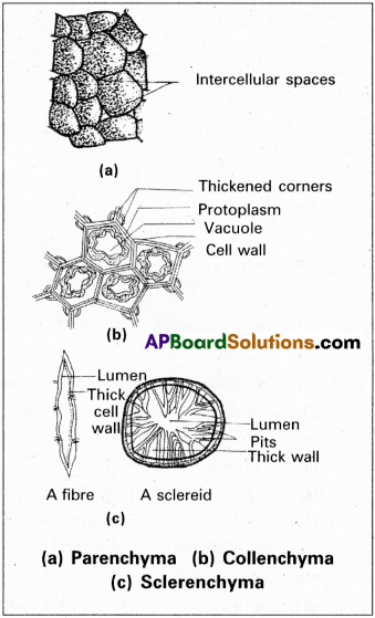
1) Parenchyma :
It is living tissue. It occupies the major part of the plant body and the cells are isodiametric. They may be spherical, oval, polygonal or elongated in shape. Their walls are thin and made up of cellulose. They have small intercellular spaces. It performs photosynthesis, storage, secretion, Healing of wounds secretion, Buoyancy by storing air etc.
2) Collenchyma :
It is simple living mechanical tissue, occurs below the epidermis of Dicot plants. It consists of cells which are thickened at the corners due to deposition of cellulose, hemicellulose and pectin. The cells are oval or spherical or polygonal in shape and contain chloroplasts. Intercellular spaces are absent. They may be angular or lacunar or Lemellar type. They help in assimilation and provide mechanical support to young stems and petiole of a leaf.
3) Sclerenchyma :
It is a dead Mechanical tissue, consists of long, narrow cells with thick and lignified cell walls having pits. They are usually dead cells and without protoplasts. Based on the form, structure, it may be either fibres or sclereids. The fibres are thick walled, elongated and pointed cells, gives mechanical support and also used in Jute Industries. The sclereids are spherical, oval or cylindrical, highly thickened dead cells, found in the fruit walls of nuts, pulp of fruits like guava and sapota, seed coats of legumes and leaves of tea. They also provide Mechanical support to organs.
Question 4.
What are complex tissues? Describe various types of complex tissues
Answer:
Tissues which are made up of more than one type of cells and work together as a unit are called complex tissues. They are of two types namely xylem and phloem.
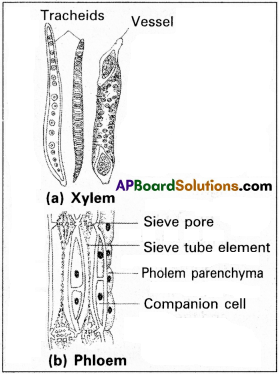
Xylem :
It is a conducting tissue, helps in conduction of water and minerals form roots to stem and leaves. It is composed of four elements namely tracheids, vessels, xylem fibres and xylem parenchyma.
Tracheids are elongated or tube like cells with thick and lignified walls and tapered ends. These are dead and are without protoplasm. Vessels are long cylindrical tube like structures, made of many cells with lignified walls and a large central cavity. Vessels are interconnected through perforations in their walls. They are, also dead cells. Both Tracheids and vessels are the main water transporting channels. Xylem fibres are long, with thick walls and narrow lumens, gives mechanial strength, xylem parenchyma cells are living and thin walled cells, made up of cellulose. They store food materials in the form of starch of fat.
Phloem :
It is a complex tissue, helps in conduction of food materials. It is composed of sieve tube elements, companion cells, phloem parenchyma and phloem fibres. Sieve tube elements are long tube like structures and are associated with companion cells. Their end walls are performted in a sieve like manner to form the sieve plates. A mature sieve element shows a peripheral cytoplasm and a large vacuole but lacks a nucleus. The companion cells are specialised parenchymatous cells which are connected to sieve tube by pit fields the companion cells help in maintaining the pressure gradient in the sieve tubes.
Phloem parenchyma is made up of cylindrical cells which have dense cytoplasm and nucleus. The cell wall is composed of cellulose and has pits. It stores food material and other substances like resins, Latex and Mucilage. Phloem fibres are long elongated unbranched cells with pointed apices. The cell wall is thick and is made up of lignin. Phloem fibres of jute flax and hemp are used commerically.
Question 5.
Describe the internal structure of dorsiventral leaf with the help of labelled diagram.
Answer:
Transverse section of a dorsiventral leaf (dicot leaf) shows 3 important parts. They are
- Epidermis,
- Mesophyll and
- Vascular byndles.
1. Epidermis :
Epidermis is present on the both the upper surface (adaxial) and the lower surface Cabaxial) of the leaf. The epidermis present on the adaxial surface is called upper epidermis and on the abaxial surface is called lower epidermis. The epidermis is made up of one row of barrel shaped cells, which are arranged compactly without intercellular spaces. The cells are filled with vacuolated and nucleated protoplast. On outerside of the epidermis a waxy layer called Cuticle is present.
Stomata are present, more on the lower surface than on the upper surface. Each stoma is surrounded by two kidney shaped guard cells. They are chlorophyllous and regulate the opening and closing of stomata. Epidermis shows multicellular uniseriate hairs. The cells of leaf hairs are filled with water. They protect the inner tissues by absorbing the heat and prevents evaporation of water from the leaf surface. The stomata help in the gaseous exchange and also promote transpiration.
2. Mesophyll :
The ground tissue that extends between the upper and lower epidermal layers is called the mesophyll. It is composed of thin walled parenchyma with chloroplasts. It is chiefly concerned with the synthesis of carbohydrates. In dicot leaves mesophyll is differentiated into two parts namely,
i) Palisade parenchyma and
ii) Spongy parenchyma.
i) Palisade parenchyma :
Part of the mesophyll found beneath the upper epidermis is called ‘palisade tissue’. It shows elongated, columnar cells arranged in 1-3 vertical rows. Narrow intercellular spaces are present between the cells. In these cells, large numbers of chloroplasts are found nearer to the cell wall. Palisade tissue is primarily concerned with the manufacture of carbohydrates in the presence of sunlight.
ii) Spongy parenchyma :
Part of the mesophyll found towards the lower epidermis is called spongy tissue. It shows 3-5 rows of irregular shaped cells that are arranged loosely with large intercellular spaces. Some intercellular spaces present in the vicinity of the stomata are very large, forming air chambers (air cavities). In thise cells, number of chloroplasts is less. That is why the upper surface of leaf is dark green and the lower surface is light green in colour. Spongy tissue has a primary role in gaseous exchange, apart from the synthesis of food materials.
3. Vascular bundles :
Vascular bundles are extended in the mesophyll in the form of veins. They help in supplying water, mineral salts and food materials all over the leaf surface. Veins also provide mechanical strength to the leaf.
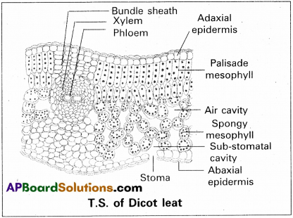
The vascular bundles are conjoint, collateral and closed. The xylem is present on the upper side and phloem on the lower side. Cambium is absent between them. Xylem shows vessles, tracheids, parenchyma and fibres. Phloem shows sieve tubes companion cells and phloem parenchyma.
Each vascular bundle is surrounded by a layer of specialised mesophyll cells that are arranged closely and compactly without intercellular spaces. This layer is called bundle sheath or border parenchyma. The bundle sheath cells divide and grow towards the upper and lower epidermal layers. These are called bundle sheath extensions. They help in the conduction of food materials form the mesophyll to the vascular bundles.
Question 6.
Describe the internal structure of an isobilateral leaf with the help of labelled diagram.
Answer:
The internal structure of a monocot leaf (isobilateral leaf) shows 3 main parts, namely 1. Epidermis, 2. Mesophyll and 3. Vascular bundles.
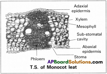
1) Epidermis :
Epidermis is present on both the upper surface (adaxial) and the lower surface (obaxial) of the leaf. The epidermis is made up of barrel shaped cells which are arranged compactly without intercellular spaces. The cells are filled with vacuolated cytoplasm and possess a single nucleus but chloroplasts are absent. Epidermal hairs are absent. Epidermis is externaly covered by a waxy layer called cuticle. The number of stomata on both the sides is almost equal. In some monocots like grasses some cells of upper epidermis are enlarged and specialised, called Bulliform cells or Motor cells. They are thin walled and are filled with water. They help in rolling and unrolling of the leaf. The epidermis gives protection to the inner tissues, regulates the transpiration and helps in gaseous exchange.
2) Mesophyll :
It is present between the upper and lower epidermal layers. It is made up of several layers of columnar cells or spongy cells, that are loosely arranged showing intercellular spaces. They contain chloroplasts. The mesophyll is the cheief photosynthetic tissue of the leaf.
3) Vascular bundles :
Numerous vascular bundles are present in the mesophyll in the form of veins. The vascular bundles are conjoint, collateral and closed. Xylem is present towards upper side and phloem towards lower side.
Each vascular bundle is enclosed by a layer of specialised mesophyll cells called border parenchyma or bundle sheath. Sometimes, bundle sheath is composed of dead sclerenchymatous tissue. The bundle sheath cells divide and grow towards both the sides of the vascular bundle. They are called bundle sheath extensions. They help in the conduction of materials from the mesophyll to the vascular bundle. They also give mechanical strength to the leaf.
Question 7.
Distinguish between the following :
a) Exarch and endarch condition of protoxylem.
b) Stele and vascular bundle.
c) Protoxylem and metaxylem.
d) Interfasicular cambium and Intrafasicular cambium.
e) Open and closed vascular bundles.
f) Stem hair and root hair.
g) Heat wood and sap wood.
h) Spring wood and Autumn wood.
Answer:
a)
| Exarch | Endarch |
| If the protoxylem lies towards periphery and metaxylem lies towards the centre is called Exarch condition. Ex : Roots. | If the Protoxylem lies towards the centre and metaxylem lies towards periphery is called Endarch condition. Ex : Stems. |
b)
| Stele | Vascular Bundle |
| Stele is the central part of the Internal structure of stems or Roots constitutes pericycle, vascular Bundles, Medulla. | Xylem and phleom are arranged on the same radius or on different radius called vascular Bundle. |
c)
| Protoxylem | Metaxylem |
| The first formed xylem with narrow lumen is called protoxylem. | The later formed xylem with broader lumen is called Metaxylem. |
d)
| Interfascicular cambium | Intrafascicular cambium |
| The cells of medullary rays adjoining intrafascicular cambium become meristematic and form interfascicular cambium. | The cambium present between xylem and phloem of a vascular bundle is called Intrafascicular cambium. |
e)
| Open Vascular Bundle | Closed Vascular Bundle |
| If cambium is present between xylem and phloem of vascular bundle then it is called open vascular bundle. Ex : Dicot stem. | If cambium is absent between xylem and phloem of a vascular bundle, then it is called closed vascular bundle. Ex : Monocot stem. |
f)
| Stem Hair | Root Hair |
| 1. They are multicellular or unicellular, separated from epidermal cells by walls. | 1. They are unicellular. They are not separated from epidermal cells by wails |
| 2. They check the rate of transpiration. | 2. They help in absorption of water from the soil. |
g)
| Heart wood | Sap wood |
| 1. The older xylem present in the centre, appears dark in colour is called Heart wood. | 1. The newly formed xylem found in the peripheral part of the plant, light in colour is called sap wood. |
| 2. It does not conduct water. | 2. It is active in conducting water. |
| 3. It is highly durable. | 3. It is less Durable and more permeable. |
| 4. It is older and harder part. | 4. It is younger and softer part. |
h)
| Spring wood | Autumn wood |
| 1. Xylem formed in springs season and have wider lumes is called spring wood. | 1. Xylem formed in autumns season with narrow lumen is called Autumn wood. |
| 2. It is light in colour. | 2. It is dark in colour. |
| 3. Formed early in a year. | 3. Formed after the early wood. |
| 4. Produced more in amount. | 4. Produced less in amount. |
| 5. Less dense. | 5. More dense. |
![]()
Question 8.
What is stomatal apparatus? Describe the structure of stomata with a labelled diagrams.
Answer:
The stomatal aparture, Guard cells and the subsidiary cells together called “Stomatal apparatus”.
Structure of Stomata :
Stomata are the structures present in the epidermis of leaves. Stomata regulate the process of transpiration and gaseous exchange. Each stoma is composed of two bean-shaped Guard cells. In grasses, the guard cells are dumbbell shaped. The outer walls of guard cells are thin and inner walls are highly thickened.
The guard cells possess chloroplasts and regulate the opening and closing of stomata. Some times, a few epidermal cells become specialised in their shape and size and are known as subsidiary cells. The stomatal aperture, guard cells and the subsidiary cells are together called stomatal apparatus.
Question 9.
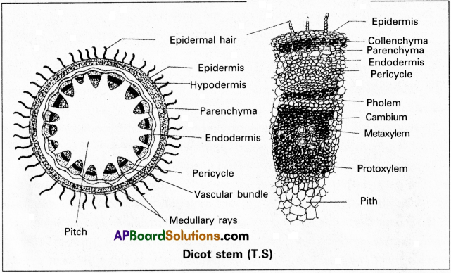
Describe the T.S of a dicot stem.
Answer:
The structure of young dicot stem can be clearly understood by observing the transverse section of stem of Helianthus annus (sunflower). It shows three major zones, namely epidermis, cortex and stele.
1. Epidermis :
It is the outer most layer of rectangular or tubular cells arranged compactly without any intercellular spaces. On outer surface of epidermis, a waxy layer called Cuticle is found. The cuticle is chemically composed of a substance cutin. The cell walls of epidermis also show the presence of cutin. Stomata are present in the epidermis. Multicellular trichomes develop on the epidermis. The cuticle and the trichomes check the evaporation of water and protect the stem from high temperature.
The epidermal layer gives protection to the inner tissues and also prevents the evaporation of water from the plant body. Through stomata, the epidermis allows the exchange of gases and promotes transpiration. Trichomes prevent entry of pathogens.
2. Cortex :
The part extending between the epidermis and the stele is known as cortex. The cortex is smaller than the stele. It shows three sub-zones, namely, i) Hypodermis ii) General Cortex and iii) Endodermis.
i) Hypodermis :
This is the outermost part of cortex and composed of 3-6 rows of collenchy matous cells. It is found beneath the epidermis and helps in providing tensile strength (elasticity) to the stem. The cells are arranged compactly without intercellular spaces and show excessively thickened corners. The cells are filled with active vacuolated cytoplasm possessing chloroplasts. Thus, the hypodermis also helps in the assimilation of food materials. It also gives mechanical strength.
ii) General Cortex :
It is found beneath the hypodermal layer and is made up of 5 – 10 rows of thin walled, living parenchyma cells with or without intercellular spaces. These cells may be isodiametric or oval or spherical. The outer layers of cells contain chloroplasts and in the inner layers leucoplasts are found. The general cortex is primarily concerned with the assimilation and storage of food materials.
iii) Endodermis :
The inner most layer of cortex is called endodermis. The cells are barrel shaped, compactly arranged without intercellular spaces. The endodermis cells contain vacuolated protoplasts and show starch grains. So it is also known as ‘Starch Sheath’.
3) Stele :
The central conducting cylinder is called the ‘Stele’. It occupies a major part of the stem. It Is composed of 4 parts.
i) Pericycle ii) Vascular bundles iii) Pith or Medulla and iv) Medullary rays.
i) Pericycle:
It is present in the form of a discontinuous ring and is made up of 3-5 rows of the thick walled, dead, lignified cells which gives mechanical strength to the stele. It appears as semilunar patches of sclerenchyma above the vascular bundles with intervening masses of parenchyma.
ii) Vascular bundles :
About 15-20 vascular bundles are arranged in the form of a ring. This arrangement is called eustele. Each vascular bundle is wedge or top shaped. In the vascular bundels xylem and phloem are arranged on the same radius. A meristematic layer of cells called cambium is present in between the xylem and phloem. So they are called conjoint, collateral, open vascular bundles. Xylem is at the lower side and phloem at the upper side of the vascular bundle.
Xylem consists of vessels and xylem parenchyma. There may befewtracheidsand xylem fibres. The metaxylem is towards the pericycle and protoxylem towards the pith. This is called endarch xylem. Phloem consists of sieve tubes companion cells and phloem parenchyma. Xylem and phloem are vascular tissues which conduct water, mineral salts and organic solutes respectively.
iii) Medulla :
It is the central part of the stele and filled with thin walled parenchymatous cells, showing intercellular spaces. It is well-developed, extensive and occupies a large part of the stele. The chief function of the medulla is to store food materials.
iv) Medullary rays :
The cells of the medulla extend to the periphery in between the vascular bundles. These cells are horizontal rows of thin walled, living and elongate radially, forming primary medullary rays.
The medullary rays connect the stele with the cortex and are helpful in lateral conduction.
Question 10.
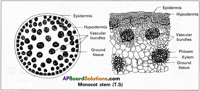
Describe the T.S of a Monocot stem.
Answer:
The structure of monocot stem can be understood well by observing the T.S of stem of Zea mays. It shows 4 distinct parts., namely
- Epidermis,
- Hypodermis,
- Ground tissue and
- Vascular bundles.
Epidermis :
It is the outermost layer composed of rectangular or tubular living cells arranged closely and compactly without intercellular spaces. The cells contain vacuolated protoplasts with a single nucleus but chloroplasts are absent. A waxy layer called ‘cuticle’ is deposited on the external surface of the epidermis. Cuticle prevents the evaporation of water from the plant body. Trichomes are absent. Numerous stomata are found in the epidermis through which exchange of gases occurs.
Epidermis gives protection to the inner tissues, helps in the exchange of gases and also prevents the evaporation of water.
Hypodermis :
A distinct cortex is absent in Monocot stems. However a thick walled hypodermis is found beneath the epidermis. The cells of hypodermis are sclerenchymatous and are arranged compactly in 3 – 4 rows, without any intercellular spaces. It gives mechanical strength to the stem.
Ground tissue :
A major part of the stem is formed by an extensive soft, parencymatous tissue called the ground tissue. The peripheral layer consists of smaller cells while the inner layers show bigger cells. The cells of peripheral layers are chlorenchymatous and are concerned with assimilation of food
Vascular bundles :
Numerous vascular bundles are found scattered irregularly in the ground tissue. This kind of arrangement is called ‘atactostele1. It is considered an on advanced character. The inner vascular bundles are bigger in size and far apart from one another. The outer vascular bundles are smaller and are close to one another and found In one or two circles.
Each vascular bundle is oval in shape and shows xylem and phloem together on the same radius. There is no cambium between xylem and phloem. Hence the vascular bundles are called conjoint, collateral and closed. Xylem is at the lower side and phloem at the upper side of the vascular bundle. Each vascular bundle is enclosed by a sheath of sclenrenchymatous fibres. Hence it is called fibro vascular bundle.
Xylem consists of tracheids, vessels, fibres and parenchyma. Xylem vessels are few in number (4) and are arranged in “Y” shape. One or two protoxylem cells are crushed forming lysigeneous cavity called protoxylem lacunae which store water phloem consists of sieve tubes and companion cells. Phloem parenchyma is absent Medulla, Medullary rays and pericycle are also absent.
Question 11.
Describe the internal structure of a Dicot Root.
Answer:
The transverse section of primary dicto root can be divided into 3 zones. They are : Epidermis, Cortex and Stele.
1) Epidermis :
Epidermis is the outermost layer made up of thin walled, non-cutinised, rectangular living cells. The epidermal cells are arranged compactly without intercellular spaces. The cuticle and stomata are absent. Some epidermal cells produce tubular extensions called root hairs. Due to presence of root hairs the root epidermis is called is as epiblema or rhizodermis or piliferous layer.
The cells that give rise to root hairs are comparatively smaller than the other cells and are called trichoblasts. Root hairs help in the absorption of capillary water. The epidermis gives protection to the inner tissues and plays major role in the absorption.
2) Cortex :
The tissue extended between epidermis and stele is called ‘cortex’. Generally in roots materials. The inner layers of cells are concerned with storage of food materials. In monocot stem endodermis is absent. the cortex is bigger than the stele. Cortex can be differentiated into three parts :
- Exodermis,
- General cortex and
- Endodermis.
i) Exodermis :
It is the outermost layer of cortex and composed of two to three rows of thick walled suberised cells. When the epidermal layer is removed, the exodermis acts as the protective layer. It also prevents the exit of water from the cortex. It can be observed in mature part of the root.
ii) General cortex :
It is present beneath the exodermis and is composed of several rows of thin walled, living parenchyma cells. The cells are round or oval in shape and loosely arranged showing intercellular spaces. They contain leucoplasts which store the food materials. The general cortex helps in the lateral conduction of water from the epidermis to the xylem vessels present in the stele.
iii) Endodermis :
It is the innermost layer of cortex and is made up of a single row of barrel shaped cells. The cells are compactly arranged without having any intercellular spaces. The radial and transverse walls of the endodermal cells show casparian strips that are formed by the deposition of lignin and suberin which prevent the movement of water. Therefore the endodermis acts as a barrier between the cortex and the stele.
In endodermis, some cells situated opposite to the protoxylem elements are thin walled without casparian bands. These cells are called passage cells. They help in the translocation of water and mineral salts from the cortex into the stele.
3) Stele :
The central conducting cylinder is known as ‘stele’. It is smaller than the cortex. The stele is comprised of three parts, viz., pericycle, Vascular bundles and Medulla.
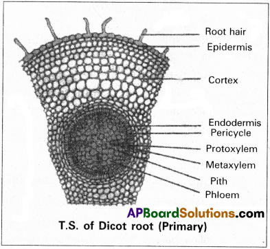
i) Pericycle :
The layer of cells surrounding the stele is known as ‘pericycle’. It is usually uniseriate and composed of thin walled, rectangular, living cells which show active cell division. The pericycle gives rise to lateral roots. Some cells of the pericycle can dedifferentiate into secondary cambium which results in the secondary growth of the root.
ii) Vascular bundles :
Standards of primary xylem and phloem are found alternately on separate radii. These are called separate or radial vascular bundles’. Xylem is exarch showing protoxylem elements towards the pericycle and metaxylem elements towards the medulla. The number of vascular bundles is identified in relation to the number of xylem groups. Usually in dicot roots, four xylem bundles alternating with four phloem bundles are found. This is called ‘tetrach condition’.
There is no cambium between the vascular tissues. The ground tissue that extends between the xylem and phloem strands is called conjunctive tissue. It is usually parenchymatous. It helps in the storage of food materials. It porduces secondary cambium during secondary growth.
Medulla or Pith :
In roots, the development of xylem is centripetal and produces metaxylem towards the inner side. Sometimes, as in dicot root these metaxylem elements come closer from all sides and replace the medulla. Hence in dicot roots the medulla is very small or may be completely absent. When present, it is parenchymatous and helps in the storage of food and water.
Question 12.
Describe the internal structure of a Monocot Root.
Answer:
The internal structure of Monocot root shows 3 zones. They are :
- Epidermis
- Cortex and
- Stele.
1) Epidermis :
It is the outermost layer formed by thin walled, rectangular cells, which are compactly arranged without intercellular spaces. Cuticle and stomata are absent. Some epidermal cells (trichoblasts) produce tubular extensions called root hairs. They absorb capillary water from the soil. The epidermis of root is also known as rhizodermis or epiblema or piliferous layer.
2) Cortex :
It is a wide and extensive tissue present between the epidermis and stele. It is bigger than the stele. It can be divided into three sub-zones. They are :
a) Exodermis
b) General cortex and
c) Endodermis.
a) Exodermis :
It is the outer part of the cortex and composed of one to two rows of thick walled, dead, suberised calls. In mature roots, when the outer epidermis is removed, the exodermis acts as a protective layer. It helps in preventing the exit of water from the root tissues.
b) General Cortex :
It is formed below the exodermis layer. It is composed of several rows of thin walled living cells that are arranged loosely showing intercellular spaces. The cells of cortex help in the storage of food materials and lateral conduction of water from the epidermis to the stele.
c) Endodermis :
The innermost layer of cortex and is composed of single layer of barrel shaped cells that are arranged compactly without intercellular spaces. The radical and transverse walls are wrapped by ligno-suberised bands called casparian bands.
Some cells situated opposite to the protoxylem cells are thin walled and without casparian bands. These are known as passage cells which help in the entry of water from the cortex into the stele.
3) Stele :
The central conducting cylinder. It is very prominent and bigger in size. The stele shows Pericycle, Vascular bundles and Medulla.
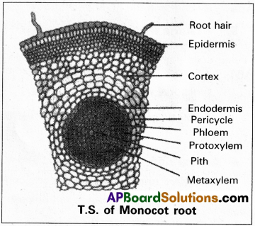
i) Pericycle :
The layer of cells found beneath the endodermis is known as pericycle. The cells are thin walled, parenchymatous, rectangular and compact without intercellular spaces. The cells are meristematic and divide actively producing lateral roots. In old and mature roots, the pericycle is sclerenchymatous and gives mechanical strength.
ii) Vascular bundles :
Bundles of xylem and phloem are found separately on different radii, one alternating with the other, at the peripheral boundary of the stele. These are known as radial’ or separate vascular bundles. The xylem is exarch and polyarch. More than six xylem bundles.
The ground tissue formed between the xylem and phloem stands is known as ‘conjunctive tissue’. It is usually parenchymatous. It helps in storage of food materials and provides mechanical strength.
iii) Medulla or Pith :
The wide central part of the stele is called ‘medulla or pith’. It is made up of thin walled parenchyma which primary helps in the storage of food. In some monocot roots, the medulla is composed of thick walled lignified dead cells and helps in giving mechanical strength.
Intext Questions
Question 1.
Name the various kinds of cell layers which constitute the bark.
Answer:
Periderm and secondary phloem.
Question 2.
Every 50 years, for 200 years, a nail was drilled into a tree, to the same depth and at exactly 1m above the soil surface (assuming the ground level has not changed). What will be the pattern of the four nails on the tree? Do you know the reason for your answer? If yes, give the reason?
Answer:
The pattern of four nails depends on the seasonal variations and growth of the tree.
Question 3.
Why is wood made of xylem and not of phloem?
Answer:
The walls of the xylem cells are thickened with lignin, this strengthens the walls and also makes them waterproof. Xylem also contributes greatly to the mechanical strength of the plant. Hence wood is mostly made up of secondary xylem.
![]()
Question 4.
A student estimated the age of a tree to be about 300 years. How did he anatomically estimate the age of this tree?
Answer:
By counting the number of annual rings.
Question 5.
Assume that you have removed the duramen part of a tree. Will the tree survive or die?
Answer:
The tree survives even if the duramen part of trunk is removed because of the presence of functional wood called sap wood.